The immune system manages a dental infection at its edges. It doesn’t clear it at the source, because the source is inside a structure with limited blood supply that prevents antibiotics and immune cells from reaching it in sufficient concentration. Without mechanical removal of the infected tissue, the infection doesn’t stabilize. It moves.
Where it moves, and how fast, follows a sequence that’s specific and well-documented. Most patients who delay treatment do so during a period when symptoms have quieted. Understanding what is happening to the tooth and the surrounding bone during that quiet period is what this blog covers.
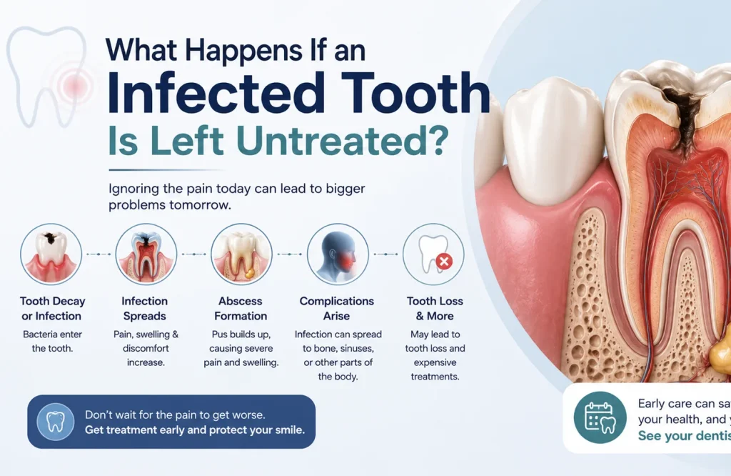
From Decay to Infection: How the Pulp Gets Involved
Bacteria reach the pulp through deep decay that has gone untreated long enough to penetrate the enamel and dentin, through a crack that provides direct access to the inner tooth structure, or through trauma that disrupts the tooth’s vascular supply without visible external damage.
The pulp chamber is an enclosed space. Blood supply enters and exits through a narrow opening at the root tip. That anatomical constraint limits what the immune system can do once infection establishes inside it. The immune response that clears most infections requires adequate blood flow to deliver white blood cells and, where antibiotics are used, sufficient drug concentration at the site. The pulp’s restricted vascularity means neither arrives in the quantities needed to clear a bacterial load that has established and is multiplying in an enclosed environment.
A skin abscess can be walled off, drained, and eventually cleared because the surrounding tissue has sufficient blood supply to support that process. The pulp chamber doesn’t have the equivalent. Inflammation builds pressure inside a sealed space, which produces the acute pain of pulpitis. The bacteria, without removal of the tissue they’ve colonized, have a single direction available to them.
When the Pain Stops, the Infection Hasn’t
Acute pulpitis produces the severe, often throbbing pain that sends patients to emergency dental appointments. That pain comes from inflamed, infected pulp tissue with an intact nerve supply. When the infection progresses to the point of complete pulp necrosis, the nerve fibers within the pulp die along with the tissue. The pain stops.
The bacteria that caused the necrosis are still present, now inside a chamber full of dead tissue that provides an ideal bacterial environment: warm, nutrient-rich, and sealed from immune activity. The infection doesn’t pause when the pain resolves. It advances through the root canal and out through the apex into the periapical bone.
Patients who experience several days of severe tooth pain followed by gradual resolution frequently conclude the infection cleared on its own. The pain cleared. The nerve that was generating it died. The infection is now progressing through bone, which has no equivalent nerve supply to signal what’s happening. That silent phase, between nerve death and the point where infection reaches soft tissue and produces new symptoms, is the period during which the most clinically significant progression occurs and during which most treatment delays happen.
Pain returns when the infection reaches structures with intact nerve supply or when abscess pressure builds sufficiently to produce symptoms at the surface. By that point, the infection is no longer within the tooth.
What Develops When Infection Reaches the Bone
Infection spreading through the root apex into surrounding bone triggers an immune response: white blood cells move to the site, pus accumulates, and a periapical abscess begins to form at the root tip. The abscess is the body’s attempt to contain bacteria it can’t eliminate. The source of those bacteria, the necrotic pulp above, is unchanged.
What happens next depends on whether the abscess finds a drainage pathway.
Without one, pressure builds. The abscess expands, erodes through bone, and migrates through soft tissue toward the surface along the path of least resistance. The pain at this stage is acute and pressure-driven: a pulsating, escalating discomfort that distinguishes an expanding abscess from the diffuse ache of earlier infection stages. Facial or gum swelling that appeared relatively quickly, feels firm and tender, and is associated with a specific tooth is the presentation most patients recognize and act on.
With a drainage pathway, the abscess forms a sinus tract: a narrow channel through bone and gum tissue that opens on the gum surface as a small pimple-like lesion. The drainage releases pressure. The pain that would otherwise have escalated doesn’t. Patients who notice the lesion often don’t connect it to the tooth; those who do frequently interpret the drainage as the infection clearing. A salty or unpleasant taste when the fistula releases is the most specific patient-detectable indicator. The infection source in the pulp is still present. The fistula is the route by which bacterial products are leaving, not the route by which the infection is resolving.
The clinical significance of the two presentations is the same. An abscess that is draining isn’t less serious than one building pressure. The pressure signal that would have driven the patient to seek care has been relieved. The infection continues at the source.
Why Antibiotics Manage the Spread but Don’t Treat the Infection
Patients prescribed antibiotics for a dental infection who experience symptom improvement within a few days frequently conclude the infection has been treated. The improvement is genuine. The infection source is unchanged.
Antibiotics reach infection sites through the bloodstream. Their effectiveness depends on blood supply delivering sufficient drug concentration to the site. A necrotic pulp chamber and an established periapical abscess both have severely restricted vascularity. Antibiotic concentration at those sites reaches a fraction of what arrives at a well-vascularized infection. Enough to reduce systemic bacterial spread and manage the acute inflammation at the infection margins. Not enough to clear the bacterial load within the necrotic tissue or the abscess cavity.
Swelling reduces. Fever comes down. The acute pain associated with spreading inflammation settles. The patient feels considerably better within two to three days of starting antibiotics. The bacteria within the necrotic pulp and the abscess are suppressed rather than eliminated. When the course ends, the source remains. The infection re-establishes from the same site, now potentially involving strains that survived the antibiotic exposure.
Repeated courses follow the same pattern: symptomatic improvement during each course, re-establishment when it ends, progressive antibiotic resistance risk with each cycle, and continued infection progression at the source level between courses. The tooth’s clinical situation worsens incrementally through each antibiotic cycle without mechanical intervention.
A GP prescribing antibiotics for an acute dental infection is managing the immediate presentation appropriately with the tools available to them. The antibiotics are functioning as intended in that context. The limitation isn’t the prescription. It’s the anatomy of the infection site, which no antibiotic reaches in the concentration required to resolve a dental infection at its source.
The Pathway From Tooth to Jaw to Neck
Most dental infections treated at the abscess stage resolve without complications beyond the tooth. Spread to the surrounding soft tissue and fascial spaces requires specific conditions: inadequate local containment, a particularly virulent bacterial strain, or delay long enough that the immune response has been overwhelmed. It isn’t the default trajectory of every untreated infection. It’s the trajectory when containment fails.
Fascial spaces are anatomical planes of loose connective tissue in the jaw, face, and neck that offer minimal resistance to bacterial spread. Infection from a specific tooth follows relatively predictable pathways based on the tooth’s position and root apex direction. Lower molar infections spread differently from upper incisor infections, and the anatomical consequences of each pathway differ in clinical significance.
Cellulitis is the first indication that infection has left the abscess and entered surrounding soft tissue. The swelling changes character: from a localized, fluctuant lump near the tooth to a firmer, less defined spreading hardness in the jaw or cheek that doesn’t point toward a drainage site. A swelling that was localized two days ago and has become a broader firmness extending into the jaw has crossed from abscess to cellulitis. The management is different because the infection is no longer contained.
From the lower jaw, infection can track to the floor of the mouth and the spaces beneath it. Bilateral spread to the submandibular, sublingual, and submental spaces produces Ludwig’s angina. The danger is anatomical: the floor of the mouth swells upward, displacing the tongue posteriorly, and the infection compresses the airway from below. Ludwig’s angina is a medical emergency not because of the bacterial load but because of where the swelling occurs. Emergency department management, including surgical drainage and airway protection, is what that situation requires.
The indicators that distinguish a spreading infection from a localized abscess are specific:
- Swelling extending beyond the tooth into the jaw, cheek, or neck
- Difficulty opening the mouth fully (trismus)
- Difficulty swallowing
- Fever above 38.5°C unresponsive to over-the-counter medication
- Difficulty breathing or a sensation of airway narrowing
Any of these warrants emergency department attendance, not a dental appointment. A localized abscess is a dental situation. These indicators mean the infection is no longer localized.
The Infection That Doesn’t Resolve Is the One That Spreads
A dental infection treated before it spreads beyond the tooth is a contained problem with a specific solution. Infected tissue removed, canals cleaned and sealed, tooth restored. The infection resolves and the tooth is saved. That outcome is available at every stage short of the point where the tooth has become unrestorable or the spread has moved beyond dental management.
What changes with delay is what the treatment has to work with. A tooth treated at the acute pulpitis stage is a different clinical situation from one treated after months of periapical bone loss. Both are treatable. The bone that has been lost, the structural integrity that has been compromised, and the complexity of the restoration that follows are all functions of how long the infection had been progressing when treatment began.
The fear most patients carry into a root canal appointment is based on a procedure that no longer exists in the form that generated the reputation. What the treatment actually involves, and why the pain most patients fear is the infection rather than the procedure, is covered in [Root Canal Myths: Is It Really as Painful as Everyone Says?](link to root canal myths blog)
An infection that has been present for weeks, or a tooth that produced pain that has since settled, warrants assessment rather than continued monitoring. A root canal consultation in Abu Dhabi establishes what is happening at the tooth level and what addressing it involves for that specific case. That picture is considerably more useful than the uncertainty of not knowing.