
Different Types of Dental Crowns Explained
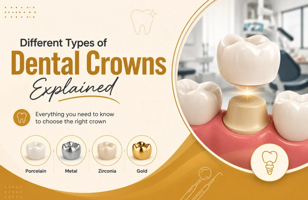
A crown recommendation usually comes with a material attached. Zirconia, E-Max, porcelain-fused-to-metal. The material is named, a price is quoted, and the conversation moves on. The clinical reasoning behind the choice rarely gets explained in the same level of detail even though understanding how crowns and bridges transform oral health helps make sense of why material matters at all. That reasoning exists and it’s specific. Crown material affects how a restoration performs under biting pressure, what it looks like against neighboring teeth, how it behaves at the gum line as tissue changes over years, and how long it holds before something goes wrong. Different materials optimize for different requirements, and the tooth’s clinical situation is what determines which requirements matter most. Understanding that logic doesn’t mean selecting the material. It means walking into the consultation with the right questions. The Four Crown Materials in Clinical Use Five crown materials are in current clinical use. What separates them isn’t a hierarchy of quality. It’s a set of different trade-offs between structural performance and optical quality, and the tooth’s clinical situation is what determines which trade-off is appropriate. Full zirconia (monolithic) is milled from a single block of zirconia with no separate substructure and no ceramic layering. The single-piece construction is the source of its strength. Earlier versions were opaque enough to limit their use to posterior positions. High-translucency zirconia has changed that, producing a material that performs well aesthetically in most positions while retaining the structural advantage of monolithic construction. Layered zirconia adds a hand-applied porcelain surface to a zirconia substructure. The substructure provides the load-bearing capacity. The porcelain layer provides the aesthetic refinement that monolithic zirconia approaches but doesn’t match in the anterior zone. The consequence of layering is that the porcelain surface becomes the point of vulnerability. Ceramic chips on layered crowns are the most common complication across all crown types that use a ceramic outer layer. E-Max (lithium disilicate) is a full glass ceramic with no substructure of any kind. Its optical properties sit closest to natural enamel of any crown material in current use. Light transmission, translucency, and color depth can be matched to adjacent teeth with a precision that no other material consistently achieves. The constraint is load: E-Max flexural strength is sufficient for anterior and premolar positions but the fracture rate in high-load molar use, particularly under parafunctional habits, is higher than zirconia alternatives. Porcelain-fused-to-metal (PFM) combines a metal substructure with a ceramic outer layer. The metal provides structural reliability. The ceramic provides tooth-colored appearance. PFM has decades of clinical data behind it and performs reliably. The issue that surfaces over time is at the margin: the metal substructure extends to the gum line, and as gingival tissue recedes naturally over years, that margin becomes visible as a dark line at the base of the crown. Full cast metal, gold or base metal alloy, has the strongest longevity data of any crown material. It requires less tooth reduction than any other crown type and tolerates the highest occlusal forces without fracture risk. The material is used exclusively in posterior positions where visibility isn’t a clinical concern, and in situations where preserving tooth structure and maximizing durability take priority over aesthetics. What Each Material Does Well and Where It Falls Short Structural performance and optical quality pull in opposite directions across all crown materials. The strongest materials are not the most aesthetically refined. The most aesthetically refined are not built for the highest forces. Where each material sits on that axis is what the clinical placement decision follows from. Full cast metal handles the highest occlusal forces of any crown material and produces no aesthetic result. Its fracture resistance under bruxism and heavy posterior load is unmatched. One specific point worth knowing: metal is softer than zirconia ceramic, which means opposing natural teeth wear less against a metal crown than against a zirconia one. For posterior positions with high functional demand and no visibility concern, that combination of properties is difficult to improve on. Monolithic zirconia is the material that handles high load with acceptable aesthetics most consistently. High-translucency versions have brought its optical quality into a range appropriate for most positions, including anterior when the aesthetic requirement isn’t the most demanding. Under bruxism conditions, it performs more reliably than any ceramic-layered alternative, with the caveat that its hardness requires careful occlusal adjustment to avoid wearing opposing natural teeth. E-Max produces the closest optical match to natural enamel of any crown material. That property makes it the default choice for anterior restorations where distinguishing the crown from adjacent teeth is the primary clinical goal. The flexural strength that works in the anterior zone is the same property that limits its use posteriorly. A patient with a heavily loaded molar, a deep bite, or significant parafunctional habits is a poor candidate for E-Max in that position regardless of the aesthetic outcome it would produce. Layered zirconia improves on monolithic zirconia’s aesthetics without fully matching E-Max. The porcelain surface layer is where the visual refinement comes from and where the complication risk concentrates. Ceramic chipping is the most frequently reported complication across all layered crown types. Patients with bruxism or heavy occlusal contacts are at meaningfully higher risk of that complication than those with lighter functional loads. In those patients, the aesthetic improvement over monolithic zirconia doesn’t justify the added vulnerability at the ceramic surface. PFM’s track record is longer than any of the ceramic alternatives. The metal substructure handles load reliably across decades. The two issues that surface over time are ceramic chipping at the biting surface and the dark line at the gum margin as tissue naturally recedes. Both are aesthetic rather than structural problems, but both are the reason patients with PFM crowns eventually return asking about replacement. Strength Aesthetics Bruxism Tolerance Best Position Full cast metal Highest None Excellent Posterior, hidden Monolithic zirconia Very high Good Very good Posterior, premolar Layered zirconia High Very good Moderate Anterior, premolar E-Max Moderate Excellent Low Anterior, premolar PFM High Moderate Moderate Variable The Tooth’s Position in the Mouth Changes the Material Priority An upper central incisor and a lower second molar require different things from a crown. The incisor sits in full view during every conversation and smile; it takes comparatively light biting forces. The molar handles the majority of chewing load and is largely out of sight. The material appropriate for one position is not the optimal choice for the other. Anterior teeth sit in what clinicians call
How to Clean Invisalign Aligners Without Damaging Them
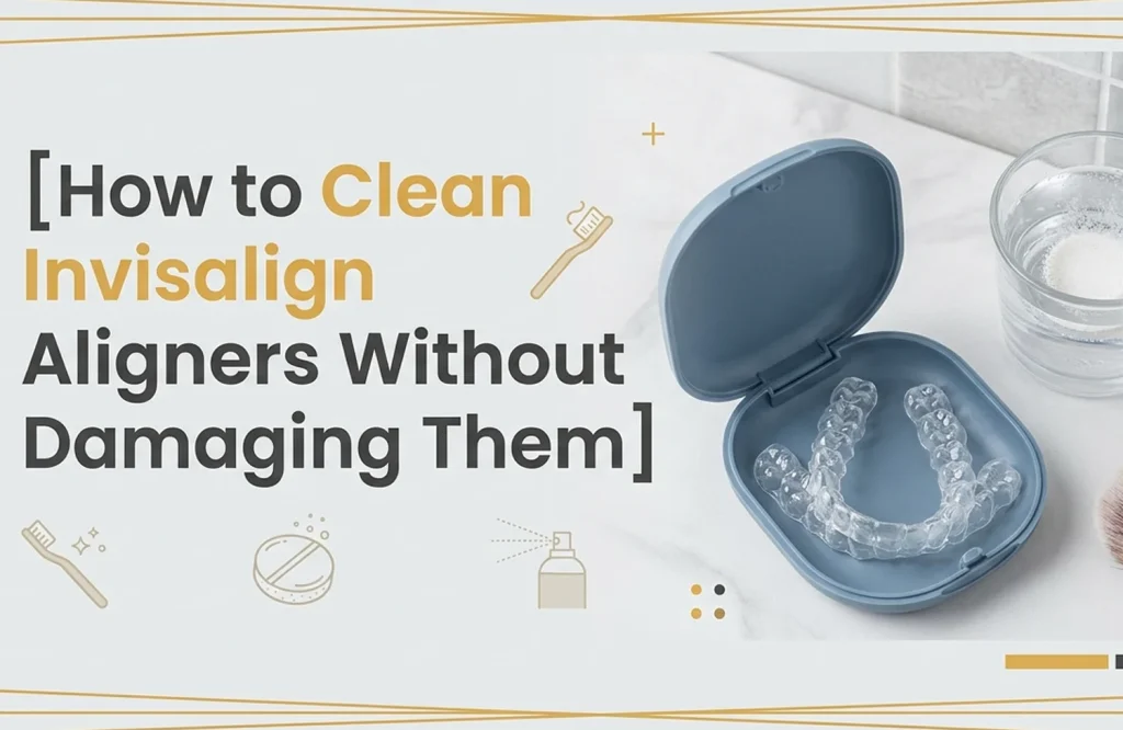
Keeping aligners clean is the part of Invisalign treatment most patients feel confident about. It seems straightforward. Rinse, brush, soak. What’s less obvious is that the cloudiness, the surface scratching, the fit that feels slightly off after a few weeks, frequently trace back to the cleaning routine rather than anything else. The plastic used in clear aligners is precise by design. It’s shaped to specific tolerances and engineered to move teeth through consistent, controlled pressure. That same precision makes the material responsive to things that seem harmless: the abrasive particles in toothpaste, water temperature, soaking solutions that weren’t formulated for thermoplastic. The damage doesn’t happen in one session. It accumulates. Understanding what the material actually responds to changes how the maintenance routine looks, and why certain habits that feel thorough are quietly working against the treatment. Why Clear Aligners Lose Their Clarity Aligners that were perfectly transparent on day one develop a milky haze within weeks. Not from neglect. From the ordinary chemistry of wearing something in your mouth for twenty-two hours a day. Saliva contains proteins that bind to the aligner surface continuously. When they’re not fully cleared between wears, they oxidize into a visible film. In Abu Dhabi, where tap water hardness runs significantly higher than WHO guidelines, mineral deposits compound that effect. Calcium and magnesium residue accumulates on the plastic surface with every rinse, dulling transparency in a way that looks like scratching but isn’t. The scratching is a separate problem. Clear aligner plastic is optically transparent because the surface is smooth at a microscopic level. Toothpaste, even varieties marketed as gentle, contains abrasive particles designed to polish enamel. Enamel is considerably harder than thermoplastic. The same particles that do nothing to a tooth surface create fine scratches across an aligner, and light scatters differently across a scratched surface than a smooth one. The result is cloudiness that no amount of cleaning reverses, because the transparency isn’t being obscured by residue. The surface itself has changed. Staining settles into that disruption. Coffee or tea consumed immediately before reinsertion, colored mouthwash used while aligners are in, pigmented foods that contact the plastic before it’s been rinsed, all of these find purchase in a compromised surface faster than they would on an intact one. The cloudier the aligner, the more aggressively it stains, and the cycle compounds. Why Some Cleaning Habits Cause More Harm Than Good The cleaning habit most reliably responsible for aligner damage is toothpaste. Not because patients are being careless with it, but because it’s sitting at the sink and it works on teeth, so the logic of using it on aligners feels sound. The abrasive compounds in toothpaste, silica in most modern formulas, are calibrated to clean enamel. Enamel has a Mohs hardness of around 5. The thermoplastic Invisalign aligners are made from sits considerably below that on the same scale. Running an abrasive designed for a harder surface across a softer one removes material from it. The effect after a single session is invisible. After two weeks of twice-daily brushing, it shows up as the cloudiness that no subsequent cleaning reverses. Water temperature produces a structurally different kind of damage. Invisalign trays are manufactured to dimensional tolerances precise enough to move specific teeth by fractions of a millimeter. The thermoplastic holds those tolerances at normal mouth and ambient temperatures. Above approximately 45 degrees Celsius, it begins to soften. The distortion from a single hot rinse is undetectable by feel. Repeated exposure changes the geometry of the tray in ways that affect how it seats against the teeth, where it applies pressure, and whether it’s still doing what it was designed to do. Patients occasionally attribute stalled tooth movement or unexpected soreness to the treatment itself. Warped trays are an underappreciated contributor to both. Soaking is where duration gets misunderstood. Retainer cleaning crystals and effervescent tablets are formulated to work within a defined window, typically fifteen to thirty minutes. The assumption that longer produces cleaner results is intuitive but wrong. Extended soaking doesn’t remove more residue. It keeps the aligner surface in prolonged contact with a chemical solution past the point it was tested for, and the cumulative effect on surface integrity across weeks of treatment is not neutral. The pressure applied during brushing matters independently of what’s being used to brush. A soft-bristled brush with a safe cleaning solution and light pressure disrupts surface deposits without abrading the plastic. Adding firm pressure to an already problematic cleaning agent accelerates surface damage at a rate the individual sessions don’t suggest. Daily Cleaning Works Best When It Stays Simple The most effective aligner maintenance routine is also the shortest. Not because shortcuts are acceptable, but because the material responds better to frequent light cleaning than to periodic intensive cleaning with the wrong products. After Removal: Saliva coats the aligner surface continuously during wear. When aligners come out and sit in open air, that film dries within minutes and becomes considerably harder to remove. A rinse under cool water immediately after removal takes under ten seconds and prevents that film from setting. It isn’t cleaning. It’s the step that makes cleaning at the end of the day less work. During the Day: Aligners removed for a meal don’t need a full cleaning cycle before going back in after plain water. After coffee, tea, or anything with color or sugar, a brief rinse and a light pass with a soft-bristled brush and no product is sufficient. The bristles disrupt surface residue without abrasion. No toothpaste. No pressure. Thirty seconds at most. The rinse that gets skipped most often is the one before reinsertion after a colored drink. That’s also the one most directly responsible for the staining that accumulates across a wear cycle and looks, by week two, like a much larger problem than it actually was at the point it could have been prevented. Before Reinsertion at Night: A fifteen to thirty minute soak in a purpose-formulated cleaning solution handles what the quick daytime rinses don’t reach. The aligner should be rinsed under cool water after soaking before it goes back in. Cleaning solution residue in contact with gum tissue across eight hours of sleep is an irritant the routine doesn’t need. Frequency matters more than intensity here. An aligner maintained at every removal stays optically clear across a full two-week cycle. The same aligner cleaned once thoroughly at the end of each day, with products that feel more rigorous than they should be, arrives at the two-week mark visibly worse. Choosing Products That Are Safe for Aligners The frame that makes this question answerable is material compatibility rather than brand preference. Invisalign aligners are made from a proprietary thermoplastic polyurethane.
Overbite vs Underbite vs Crossbite Explained
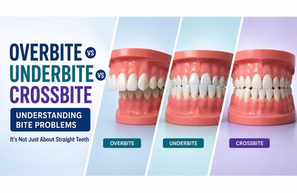
Teeth can appear straight and still have a bite problem Straight teeth don’t automatically indicate a normal bite. Alignment and bite function are related, but they are not the same thing. Teeth can appear well positioned while the upper and lower arches meet in a way that places uneven demands on the bite. Minor irregularities can also be visible without significantly affecting how the teeth function together. Overbites, underbites, and crossbites sit in that space between appearance and function. They describe changes in the way the upper and lower teeth come together rather than how straight a smile appears at first glance. Why upper and lower teeth are meant to fit together a certain way A bite isn’t simply a matter of whether teeth look straight. Every time the mouth closes, dozens of teeth come into contact in a sequence that distributes force across the entire system. The arrangement is deliberate. Front teeth guide certain movements. Back teeth absorb most of the pressure generated during chewing. Problems rarely begin because a single tooth is out of place. More often, the relationship between the upper and lower arches changes. Pressure starts concentrating where it wasn’t intended to. Some teeth begin carrying a greater share of the workload while others contribute less. Uneven force distribution is not always easy to recognise without examining how the teeth come together. A smile can appear well aligned while the bite underneath functions very differently. Understanding how the upper and lower teeth are supposed to meet makes it easier to see why overbites, underbites, and crossbites are treated as separate conditions rather than different versions of the same problem. When the upper teeth sit too far forward An overbite describes a front-to-back imbalance between the upper and lower arches. A certain amount of overlap is normal. Most upper front teeth naturally sit slightly ahead of the lower front teeth. The difference lies in the amount of overlap and the way it influences the rest of the bite. Some overbites are limited to the teeth themselves. Others are influenced by the position and development of the jaws. Two patients may both be told they have an overbite while presenting with very different underlying causes and different levels of severity. As the overlap increases, the distribution of force changes. Front teeth may absorb pressure they were never intended to handle repeatedly, while lower teeth can begin contacting areas they normally wouldn’t. In more pronounced cases, the bite stops functioning as a balanced system and starts directing pressure toward a smaller group of teeth. The effect extends beyond appearance. Tooth wear, uneven loading, and changes in chewing patterns are often linked to the way the upper and lower arches interact rather than how the teeth look when viewed from the front. When the lower teeth extend beyond the upper teeth An underbite reverses the relationship described in an overbite. Instead of the upper front teeth sitting ahead of the lower teeth, the lower arch extends further forward when the jaws close. In some cases, the difference comes primarily from tooth position. In others, the lower jaw itself sits further forward relative to the upper jaw. Similar bite patterns can arise from very different underlying structures. Jaw position often plays a much larger role in an underbite than it does in many overbite cases. Chewing changes when the front teeth no longer meet in their intended positions. Certain movements become less efficient, and some teeth begin taking on work they were not designed to handle repeatedly. Speech can also be affected, particularly when sounds rely on precise contact between the upper and lower front teeth. Wear patterns associated with an underbite tend to develop differently from those seen in an overbite. The teeth are not simply misaligned. The entire relationship between the upper and lower arches has been reversed. When teeth bite inside instead of outside A crossbite sits outside the pattern established by overbites and underbites. Those conditions describe a front-to-back imbalance. A crossbite develops across the width of the bite instead. The difference can involve a single tooth or several teeth. It may appear on one side of the mouth or across both arches. Regardless of extent, the relationship changes in the same direction. Teeth that would normally sit slightly outside their opposing teeth begin meeting on the inside. Because the imbalance occurs across a different plane, crossbites are evaluated differently from other bite problems. The question is no longer how far forward or backward the arches sit, but how they relate side to side when the jaws close. Why these bite problems affect more than appearance The visible part of a bite problem is usually what attracts attention first. Upper teeth may sit further forward than expected, the lower jaw may appear more prominent, or a group of teeth may meet in an unusual position. Those changes are easy to notice because they affect appearance. The functional side develops more quietly. Teeth are designed to share force with neighbouring teeth and opposing teeth. When the relationship changes, that workload becomes less evenly distributed. One area absorbs more pressure. Another contributes less. The difference may be small at first, but chewing happens thousands of times every week. For that reason, orthodontists don’t evaluate overbites, underbites, and crossbites as cosmetic variations of the same issue. The position of the teeth matters, but so does the way the entire system works once the mouth closes. How orthodontists determine which bite problem is present Appearance is only one part of the picture. Two patients may present with a similar looking overbite while arriving there for completely different reasons. In one case the teeth are responsible for most of the discrepancy. In another, the position of the jaws is driving the relationship. Photographs and digital scans help document what is visible. The more important question is often what happens when the teeth come together. Contact points throughout the bite, the position of the jaws, and the way the arches relate to
Habits That Help Maintain Whitening Results Longer

Whitening aftercare advice tends to arrive as a list with no priority order. Avoid coffee. Use whitening toothpaste. Rinse after meals. The individual items aren’t inaccurate. The format implies they’re roughly equivalent in impact, which they aren’t. A patient who can only realistically change a few habits needs to know which few habits actually affect how long the result holds. The two-week post-treatment window is its own specific period, covered in detail in How Long Does Professional Teeth Whitening Last? Past that window, maintenance looks different. The habits that carry the most weight in the long-term picture aren’t always the most prominent ones on the standard list, and the clinical reason behind each one is what makes it worth following rather than ignoring. Brushing Frequency and Timing Matter More Than Technique Twice-daily brushing is the baseline for oral health generally. For whitening maintenance specifically, when those sessions happen matters more than the fact of them. A patient who brushes at 7am and 10pm and drinks three cups of coffee in between is leaving chromogenic compounds in contact with enamel for hours at a stretch. The brushing frequency is correct. The maintenance value of it is undermined by the timing. Chromogenic compounds from coffee, tea, and dark foods begin binding to enamel within minutes of contact. Saliva clears some of that passively during waking hours, but the rate is insufficient when exposure is repeated across a day. Mechanical disruption is what actually interrupts the binding process. Brushing after the last coffee of the day rather than two hours later isn’t a significant change in routine. The chromogen contact time it eliminates across a year of daily coffee consumption is. The pre-sleep session carries weight independently of chromogen timing. Salivary flow drops significantly during sleep, removing the passive clearance that operates during waking hours. Whatever surface deposits are present at bedtime have six to eight hours of minimal salivary activity ahead of them. A fluoride toothpaste at that session supports enamel remineralization overnight rather than just clearing the surface. One specific qualifier: after acidic food or drink, waiting thirty minutes before brushing is clinically appropriate because acid temporarily softens enamel and brushing immediately causes surface abrasion. After non-acidic chromogenic drinks, including most coffees and teas, that rationale is weaker than the contact-time argument. The practical guidance is to brush after the coffee. The waiting logic applies to acid, not to chromogens. Reducing Staining From Diet Without Eliminating It Telling a patient who drinks two cups of coffee every morning to avoid coffee is advice that gets filed and forgotten. For the full list of foods that stain your teeth, the useful question here is which chromogenic sources stain fastest, what determines that rate, and what practical interventions reduce exposure without requiring dietary restructuring. Red wine sits at the top of the staining rate hierarchy for most patients, and the reason goes beyond its chromogen concentration. The acidity temporarily increases enamel porosity at the point of contact, giving those compounds structural access that a non-acidic chromogen source of equivalent concentration wouldn’t have. The combination of chromogenic content and acid is what makes red wine a more potent staining agent than its color alone would suggest. Coffee stains faster than most patients expect relative to tea. Temperature is a significant contributor. Hot liquid temporarily increases enamel permeability, delivering chromogenic compounds to a transiently more permeable surface than the same drink consumed cold. A hot coffee consumed quickly is a different enamel exposure event than an iced coffee of identical composition. Within the tea category, black tea stains faster than green because of higher tannin concentration. Herbal teas without tannins are largely neutral regardless of color. Drinking through a straw is a commonly cited tip that works under specific conditions. For cold drinks consumed through a straw positioned toward the back of the mouth, direct enamel contact is meaningfully reduced. For hot drinks consumed through a straw held near the lips, the practical reduction is minimal. The straw guidance applies most usefully to cold brew coffee, iced tea, and cold chromogenic drinks rather than the hot beverages that represent most patients’ primary daily exposure. Rinsing with water immediately after consuming a chromogenic drink is more effective than it sounds and requires nothing. Water reduces the concentration of chromogens remaining on the tooth surface and accelerates salivary clearance of what’s left. A rinse within two minutes of finishing a coffee leaves a lower residual chromogen load on the enamel surface than waiting until the next brushing session. The mechanism is simple and the habit adds under thirty seconds to a routine that already involves finishing a drink. Consuming chromogenic drinks as part of a meal rather than between meals changes the exposure profile in a way most patients don’t account for. Eating stimulates saliva production significantly above the resting rate. That elevated salivary flow during a meal clears chromogenic compounds from the enamel surface passively in a way that standalone drink consumption between meals doesn’t generate. The same coffee consumed with breakfast clears faster than the same coffee two hours later. No product involved, no dietary change required. What Whitening Toothpaste and Mouthwash Actually Do Whitening Toothpaste: Whitening toothpaste removes extrinsic surface deposits through mild abrasion before they embed into the enamel surface. A tooth surface carrying less chromogenic deposit restains more slowly than one carrying more, so the contribution to maintenance is real. The boundary of that contribution is also specific: whitening toothpaste doesn’t penetrate enamel and doesn’t maintain the internal color change professional whitening produced. The shade improvement from treatment happened below the enamel surface. No toothpaste reaches that layer. The abrasive level in the formula matters more than the whitening claim on the packaging. Relative Dentin Abrasivity, the RDA score, measures how abrasive a toothpaste is against tooth structure. High-RDA whitening formulas used daily cause enamel surface wear that increases porosity over time. More porous enamel takes up chromogenic compounds faster than intact enamel. A high-RDA whitening toothpaste used consistently in the belief that it protects the result can produce the opposite effect at the surface level across months of daily use. A lower-RDA formula with fluoride is a more defensible maintenance choice. Fluoride supports enamel remineralization, which reduces chromogenic uptake indirectly but measurably. Mouthwash: Alcohol-containing mouthwash is the product most likely to work against whitening maintenance without the patient recognizing it as the source. Alcohol is a desiccant. It temporarily reduces salivary flow and increases enamel surface porosity after use. A patient finishing their oral hygiene routine with an alcohol-containing rinse is ending
How Long Does Professional Teeth Whitening Last?
Professional teeth whitening results typically last between one and three years. That range is accurate and almost entirely useless without context for what sits at each end of it. Two patients treated on the same day with the same protocol can look significantly different at the six-month mark. One holds close to the result they left the clinic with. The other has faded noticeably. The treatment was identical. What differed was a combination of clinical factors present before the appointment and daily habits in the weeks after it. The variables that determine where a patient lands within that range are mostly knowable in advance. Understanding them produces a more specific expectation than a number that spans two years. Why Whitening Results Don’t Stay Fixed Whitening works by driving peroxide into the enamel and dentin, where it oxidizes the pigment molecules responsible for discoloration. The result is a chemical change in how the tooth absorbs and reflects light. The pigment molecules that remain after treatment, and the ones that enter the tooth from diet and lifestyle afterward, gradually shift the shade back over time. That’s the baseline mechanism of fading, and it’s slow. What patients notice first is usually something else. Peroxide temporarily increases enamel porosity and draws moisture from the tooth during treatment. Dehydrated enamel scatters light differently than hydrated enamel, producing a shade that appears whiter than the tooth’s stable post-treatment color. Over the following days, as the tooth rehydrates, that shade settles. The result at day seven looks different from day one, and patients who track this closely often conclude the whitening is already reversing. The rehydration process is completing, not the result failing. The stable shade at seven to ten days post-treatment is the actual outcome. Anything measured before that is the dehydration effect. After rehydration stabilizes, fading is driven by chromogenic compounds from diet and lifestyle re-entering the enamel tubules. Coffee, tea, red wine, dark sauces, tobacco; the rate at which these compounds accumulate depends on enamel porosity, consumption frequency, and contact time before they’re cleared by saliva or brushing. That accumulation, building gradually across months, is what most patients experience as their result fading. The shade immediately after whitening reflects dehydration, not the final result. The stable post-treatment color settles at around seven to ten days. The Habits and Factors That Accelerate Fading Fading speed varies considerably between patients, and most of that variation is explainable before treatment ends. The variables split into two categories: the ones a patient influences daily and the ones established by their tooth structure before the first appointment. Dietary chromogens drive restaining for most patients. Coffee and tea are the most consistent contributors, not because of acidity but because of tannins and other chromogenic compounds that bind to enamel on contact. A patient drinking three cups of coffee daily without rinsing afterward is presenting those compounds to the enamel surface multiple times across every day. Red wine, dark berries, tomato-based sauces, and soy sauce operate on the same principle at varying concentrations. Tobacco accelerates restaining beyond dietary sources. Nicotine and tar compounds penetrate enamel and accumulate in a way that surface cleaning doesn’t address between whitening cycles. The character of tobacco-related discoloration is also less predictable on repeat treatment than dietary staining, which means the longevity picture for a patient who smokes is structurally different from one who doesn’t. Acidic food and drink erodes enamel surface over time independently of what chromogens are consumed alongside it. A more porous enamel surface gives chromogenic compounds greater structural access, which is why patients with high acid diets tend to restain faster than their chromogen intake alone would predict. The first two weeks after treatment carry more weight than the months that follow them. Enamel porosity is elevated during the post-treatment period. Chromogenic compounds bind more readily to that surface than they will once it stabilizes. A patient who returns to unrestricted coffee and tea consumption within the first few days isn’t just affecting the immediate result. They’re establishing a lower baseline from which all subsequent fading occurs. Two structural factors sit outside daily habits entirely. Individual enamel density varies between patients at a baseline level that no post-treatment protocol changes. Some teeth are structurally more permeable to chromogenic uptake than others regardless of diet. Underlying dentin shade sets a ceiling on both how bright the result can be and how stable that result holds over time. Darker baseline dentin tends to produce results that are harder to achieve and less stable in the longer term. Both factors are worth understanding before the appointment rather than after. What the Post-Treatment Period Actually Requires The first two weeks after whitening have a larger effect on how long the result holds than any equivalent two-week period in the months that follow. Enamel porosity is elevated from the peroxide process. The decisions made during that window don’t just affect the immediate shade. They establish the baseline from which all subsequent color change accumulates. The most effective guidance for that period is also the least dramatic — see what to eat after whitening for the specifics. Chromogenic food and drink avoided during the first two weeks isn’t a permanent dietary restriction. It’s a targeted reduction in exposure during the window when enamel is most susceptible to uptake. Drinking coffee or tea through a straw reduces direct surface contact without requiring abstinence. Rinsing with water after staining foods reduces the contact time of chromogenic compounds before they bind. Neither intervention is significant in isolation. Across fourteen days of elevated enamel porosity, the cumulative reduction in chromogenic exposure meaningfully affects the result that stabilizes at the end of that period. Professional cleaning before whitening is worth mentioning because patients who skip it often don’t realize what it contributes. A scale and polish removes the surface deposit layer that would otherwise reduce peroxide penetration and produce an uneven result. The same whitening treatment on a clean enamel surface versus one carrying months of surface accumulation starts from a fundamentally different baseline. That difference shows up in both the immediate result and how stable it is in the months following. Whitening toothpaste does something specific and limited in the maintenance period. The mild abrasives it contains address extrinsic surface deposits before they embed. They don’t penetrate enamel and don’t maintain the internal color change the whitening procedure produced. Used consistently, whitening toothpaste keeps the surface cleaner between treatments. It doesn’t extend the treatment result itself, and patients who expect it to are working with an inaccurate model of what the product does.
Does a Root Canal Hurt? | Marigold Dental UAE
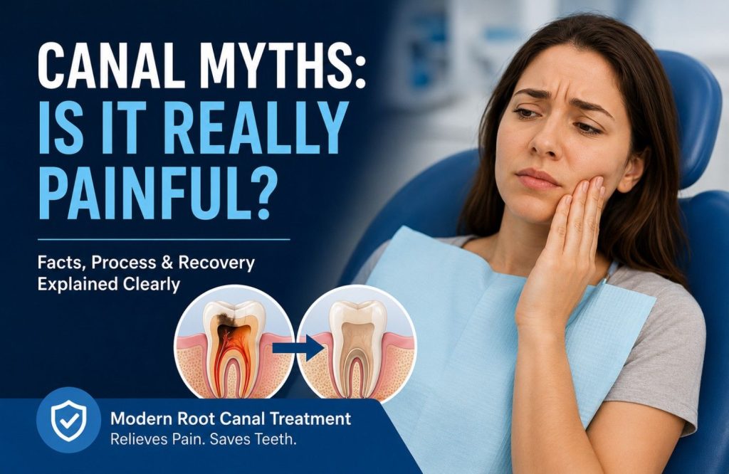
Few dental procedures carry the kind of reputation a root canal does. It’s become shorthand for something deeply unpleasant, referenced in conversations that have nothing to do with dentistry. Most patients have formed an expectation of the appointment long before they’ve been told they need one. That reputation has a history, and the history is real. Root canal treatment decades ago was a different procedure. Less precise instruments, less reliable anesthesia, longer appointments. The experience people describe, or the experience their parents described to them, came from that era. The clinical reality has moved considerably since then. The cultural memory hasn’t. What rarely gets addressed clearly is where the pain actually originates. The severe, throbbing ache that brings most patients to the clinic is the infection inside the tooth, not something the procedure creates. The root canal removes the source of that pain. That distinction is where most of the fear falls apart under examination. Why Root Canals Got That Reputation in the First Place Root canal treatment decades ago was a genuinely different procedure, and that’s where the reputation starts. Local anesthetics were less refined. Delivery techniques were less precise. Achieving full, reliable numbness in a tooth with an active infection was a real clinical limitation, not a failure of effort. Patients felt things during procedures that patients today typically don’t, and root canals, which involve working at the level of the nerve, sat near the top of that list. The instruments were different too. Manual files worked through narrow, curved canals slowly and with significant chair time. Modern rotary instrumentation uses flexible nickel-titanium files driven by a motor, faster, more precise, and considerably less demanding on the patient. Digital imaging now maps the root canal anatomy in three dimensions before anything begins. Curved roots, additional canals, unusual anatomy, all of it known in advance rather than encountered mid-procedure. The clinical experience has moved. The cultural memory hasn’t. Someone who had a root canal in the 1980s described it to someone who described it to someone else, and those accounts have been reinforced by decades of passing references in conversation, television, and film. The procedure being described in most of those accounts doesn’t exist in the same form. The fear it generated has proven considerably more durable than the techniques that caused it. The Infection Is the Problem. The Procedure Is the Solution. Beneath the enamel and dentin sits a soft inner tissue called the pulp, containing the tooth’s nerves and blood supply. In a healthy tooth it goes unnoticed. When bacteria reach it through deep decay, a crack, or trauma, that changes quickly. Infected pulp becomes inflamed inside a sealed chamber with no outlet for the pressure building within it. That pressure is what produces the severe, often throbbing pain that characterizes a tooth in need of root canal treatment. It doesn’t respond predictably to over-the-counter medication. It doesn’t resolve on its own. It progresses. As the infection advances beyond the root tip into surrounding bone and tissue, a periapical abscess develops. The pain shifts character, becoming deeper, more constant, and in some cases accompanied by swelling that spreads into the jaw. A root canal removes the infected pulp, cleans and disinfects the canal system, and seals it. The source of the pain is eliminated. What follows in the days after treatment is soreness in the surrounding tissue as it settles, not comparable in character or intensity to what preceded the procedure. Most patients fear what they’re already experiencing. The treatment resolves it. What Happens During the Appointment, Step by Step Local anesthesia comes first. The tooth and surrounding tissue are fully numb before anything else begins. For most patients the injection is the sharpest sensation of the entire appointment, and it’s the same injection given before a filling. Once the tooth is numb, a rubber dam is placed around it. A thin sheet of material that isolates the tooth, keeps the field dry, and gives the dentist a clean working environment. It’s routine, and most patients forget it’s there within a few minutes. A small opening through the crown of the tooth gives access to the pulp chamber. Fine instruments called files remove the infected tissue and shape the canal walls from inside. The canals are irrigated throughout with an antimicrobial solution, clearing bacteria and debris as the work progresses. When the canals are clean and dry, they’re filled with gutta-percha, a material that seals the space and prevents bacteria from re-entering, and the access point is closed. A crown typically follows at a separate appointment. Root canal treated teeth lose their internal moisture source and become more brittle over time. On a molar taking the full force of chewing, a crown isn’t optional. On a front tooth, the clinical picture determines whether one is needed. One to two appointments covers most cases. More complex root anatomy or a severe infection going in can extend that. During the Procedure, After It, and What’s Normal in Between With effective local anesthesia, what registers during a root canal is pressure and movement, not pain. The tooth is numb, the surrounding tissue is numb, and the sensation of instruments working inside the canal is present but not acute. Some patients are surprised by how little they feel. Others find the pressure uncomfortable even without pain, particularly during deeper canal work. Both are normal responses to the same procedure. One situation is worth addressing directly. A tooth with an acute, actively spreading infection can resist local anesthesia more than a healthy tooth does. Inflamed tissue has a lower pH, which affects how the anesthetic binds. An experienced clinician recognizes this, uses supplemental injections or alternative delivery points, and doesn’t proceed until the tooth is adequately numb. That’s not a negotiable step. In the Hours After As anesthesia clears, typically two to four hours after the appointment, mild to moderate soreness in the tooth and surrounding tissue is expected. The area has been worked, and the tissue around the root tip responds to that. Ibuprofen or paracetamol manages it in most cases. Some patients need neither. Soreness tends to peak within the first 24 hours. The tooth may feel sensitive to pressure or touch in that window; avoiding chewing on that side for the remainder of the day is the practical response. The Days Following By the second or third day the soreness is usually settling. Some tenderness when biting can persist for up to a week as the tissue around
Early Signs of Gum Disease to Watch
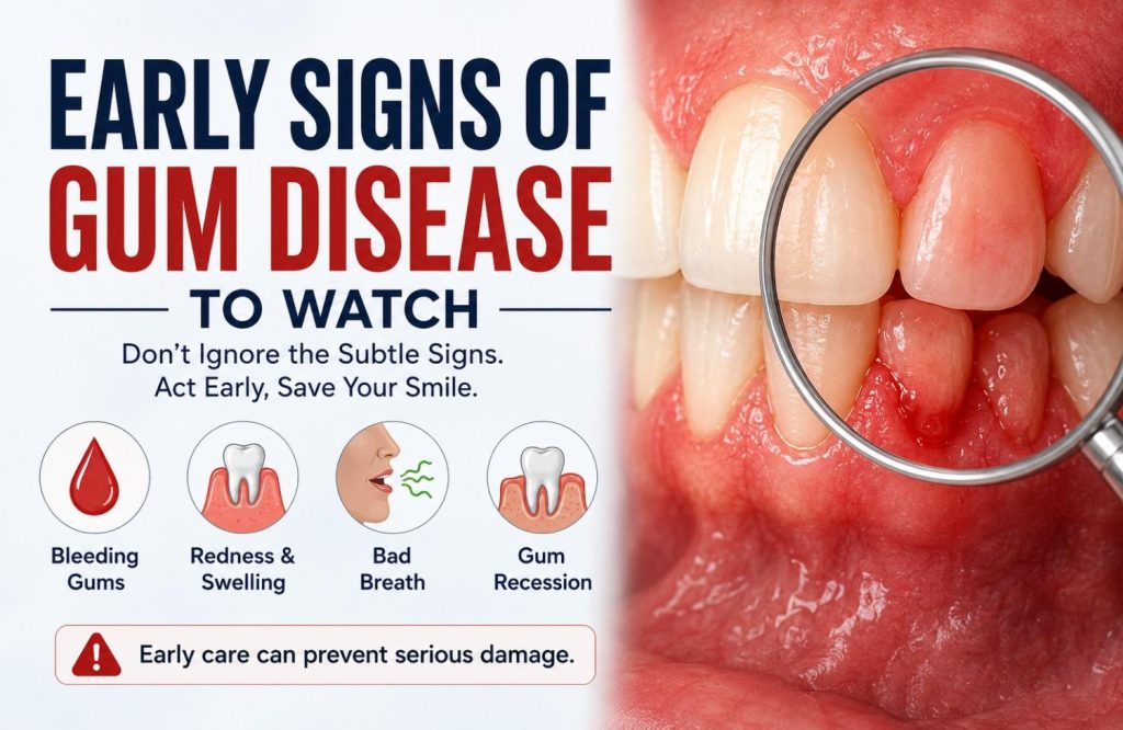
Most dental problems announce themselves. A toothache, a cracked filling, an abscess; these things produce pain that eventually becomes impossible to ignore. Gum disease doesn’t follow that pattern. In its early stages, the stages where treatment is most straightforward and outcomes are most complete, it produces signs that are genuinely easy to dismiss. Bleeding when brushing gets attributed to technique. Puffiness along the gum line is assumed to be temporary. Bad breath gets managed with mouthwash rather than investigated. None of these explanations are unreasonable. They’re just frequently wrong. The difficulty with gum disease isn’t that the signs are invisible. It’s that they’re easy to rationalize away before they’ve been properly assessed. By the time the condition causes discomfort or visible damage, it has usually been developing quietly for considerably longer. Why Most People Don’t Notice It Until It’s Further Along Plaque, the soft bacterial film that forms on teeth throughout the day, accumulates along and beneath the gum line when it isn’t consistently removed. Left undisturbed long enough, it hardens into calculus that brushing can’t clear. The bacteria within that deposit trigger an inflammatory response in the surrounding gum tissue. That’s where gum disease starts. In its earliest form, the inflammation is confined to the soft tissue. Bone and connective tissue aren’t involved yet. The signs it produces are subtle: slight redness, some puffiness, bleeding when the gums are brushed or probed. Subtle enough that most patients either don’t notice or notice and assume it will settle. What separates gum disease from most dental problems is the nature of what it destroys. Decay reaches the nerve and produces pain that eventually becomes impossible to ignore. Gum disease breaks down the bone and connective tissue supporting the tooth gradually, and those structures don’t carry the same nerve supply that signals acute pain. Significant bone loss can occur over months or years without the patient feeling it happen. The absence of pain is not evidence that the condition is stable or absent. It’s a feature of how the disease progresses, and it’s the main reason patients arrive at a more advanced stage than they needed to. What the Signs Actually Look Like Before It Becomes Obvious Each of the following signs has a plausible innocent explanation most people apply automatically. That’s exactly why they’re worth examining more carefully. Bleeding gums during brushing or flossing. Healthy gums don’t bleed in response to normal cleaning regardless of brushing pressure. When gums bleed consistently, even occasionally, it’s the tissue responding to bacterial inflammation beneath the gum line, not a technique problem. The frequency is what matters: a one-off episode is different from something that happens reliably. Redness or a purple tinge along the gum margin. Healthy gum tissue is a firm coral pink and fits tightly against the tooth surface. Tissue that looks redder, darker, or more saturated reflects increased blood flow to chronically inflamed gums. Swollen or puffy gum margins. A healthy gum margin has a crisp, tight edge where it meets the tooth. Inflamed tissue loses that tightness, becoming rounded and edematous. The change is subtle enough to miss unless you’re looking for it. Tenderness along the gum line. Not sharp pain, a sensitivity to touch or pressure along the margin that wasn’t there before. It indicates active inflammation in the tissue before it has progressed to deeper structures. Gum recession. The gum line pulling back from the crown of the tooth, exposing more of the root surface than was previously visible. Recession often happens gradually enough that it goes unnoticed until one tooth looks noticeably longer than its neighbors. Persistent bad breath that brushing doesn’t resolve. Bacterial activity within periodontal pockets produces volatile sulphur compounds. Mouthwash reduces the odor temporarily. It doesn’t reach the source. A recurring bad taste with no obvious cause. Often connected to the same bacterial activity, particularly when a pocket has become more actively infected. Bleeding gets attributed to a new toothbrush. Recession gets attributed to age. Bad breath gets attributed to diet. None of these explanations are unreasonable on their own. They become a problem when they’re used to avoid an assessment that would settle the question in a single appointment. The Difference Between Gingivitis and Periodontitis The stage gum disease is at determines what treatment looks like and what outcome is realistic. That distinction matters more than most patients realize when they’re first told they have it. Gingivitis is inflammation confined to the gum tissue. Bone and connective tissue anchoring the tooth aren’t involved yet. At this stage the damage is reversible. Professional cleaning to remove the calculus and plaque driving the inflammation, combined with consistent home care, typically returns the tissue to full health. No bone has been lost. The condition, caught here, is fully resolvable. Periodontitis is what gingivitis becomes without intervention. The infection spreads below the gum line into bone and connective tissue. Periodontal pockets deepen as tissue is destroyed, creating a sheltered environment where bacteria accumulate and the condition progresses further. Bone lost to periodontitis doesn’t fully regenerate. The disease is managed rather than reversed, and managed well it remains highly stable, but the baseline is permanently lower than it would have been at the gingivitis stage. The shift between the two doesn’t produce a noticeable signal. No new symptom, no pain, no visible change that marks the transition. It happens below the gum line over a timeline that varies considerably between patients. Immune response, genetics, smoking, systemic health, and oral hygiene habits all influence how quickly or slowly the progression occurs. The only way to know which side of that line you’re on is a clinical assessment with probing measurements and X-rays. The difference between gingivitis and periodontitis is the difference between reversible and irreversible. That distinction is what makes the timing of assessment matter. The Systemic Connection Most Patients Aren’t Told About Periodontal disease is classified as a dental condition. Its consequences extend well beyond the mouth. Inflamed gum tissue at the pocket lining is ulcerated, which gives bacteria and bacterial byproducts a direct route into the bloodstream. In a healthy mouth that pathway is largely closed. In a mouth with active periodontitis it’s continuously open. The cardiovascular link is the most extensively researched. People with periodontitis carry a statistically higher risk of heart disease and stroke. The mechanisms proposed include direct bacterial seeding of arterial tissue and the chronic systemic inflammatory state that periodontal infection sustains contributing to atherosclerosis. The relationship is substantial enough that cardiologists and periodontists increasingly treat the two conditions as connected rather than parallel. The relationship with diabetes runs in both directions. Poorly controlled blood sugar impairs the immune response to periodontal bacteria, making diabetic patients more susceptible to gum disease and slower to respond to treatment. Active periodontal infection simultaneously
Dental Implants vs Dentures Compared
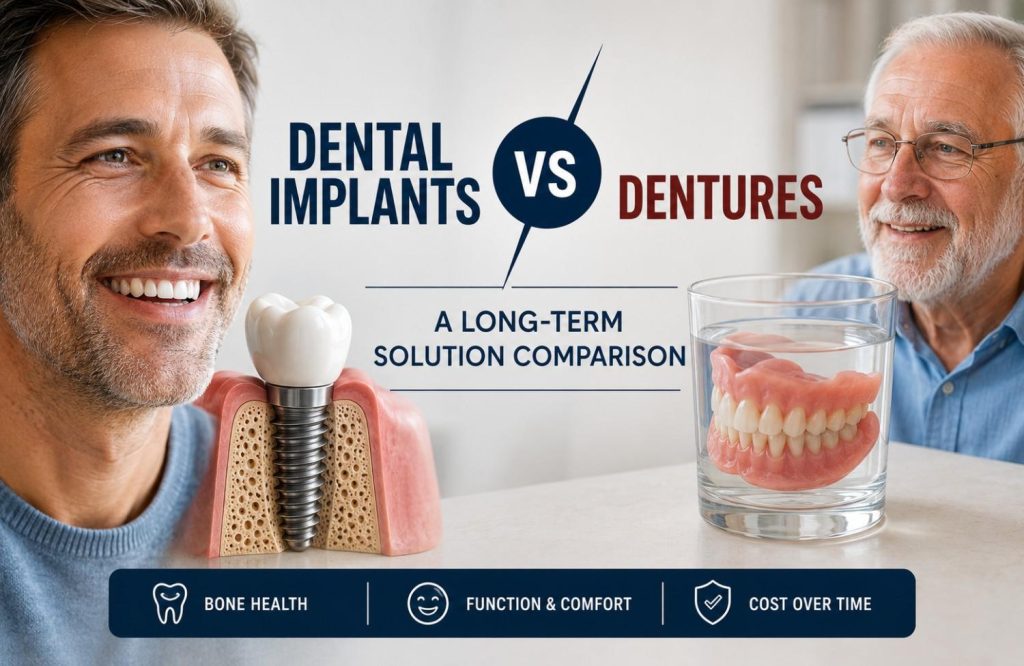
Replacing a missing tooth is a decision most people make once and live with for years. The surface comparison between implants and dentures is easy enough: one costs more, one involves surgery, one comes out at night. Most accounts of the two options don’t go much further than that. The upfront difference is only part of what separates them. What each option does to the jaw over time, how each performs under the daily demands of eating and speaking, what each actually costs across ten or twenty years rather than at the point of treatment, these are the criteria that change how the decision looks. Neither option is universally better. Which one is right depends on clinical candidacy, long-term goals, and a financial picture that goes beyond the first invoice. That’s the comparison this blog covers. Two Different Approaches to the Same Problem A dental implant is a titanium post placed surgically into the jawbone. Over the months following placement, the bone grows around and fuses with the post, a process called osseointegration. An abutment and crown are then attached on top. The finished result is fixed, non-removable, and anchored in the jaw the same way a natural tooth root is. It takes biting pressure directly. It doesn’t shift, require adhesive, or need to be removed. This is part of why implants are a long-term solution for many patients. Dentures are removable prosthetics that rest on the gum tissue surface. A full denture replaces all teeth on an arch, held in place by suction against the gum and palate. A partial denture replaces some teeth and uses clasps on remaining natural teeth for additional retention. Neither type connects to the bone beneath. They sit above it, supported by tissue that changes shape over time. For a closer look at the types and benefits of dentures, see our full guide. The distinction between embedded in bone and resting on tissue is what produces the differences in bone health, stability, and long-term function that the rest of this comparison covers. Dental Implants Dentures Placement Surgically embedded in jawbone Rests on gum tissue surface Removability Fixed, non-removable Removable Bone integration Fuses with jawbone over time No integration with bone Support structure Jawbone Gum tissue, suction, or clasps Function Comparable to natural tooth Prosthetic appliance The Bone Health Question Neither Option Can Ignore The jawbone maintains its density through stimulation. In a natural tooth, that stimulation comes from the root transmitting biting forces into the surrounding bone with every chew. When a tooth is lost, the stimulation stops. The bone in that area begins to resorb, losing volume progressively. It’s a slow process and largely invisible in its early stages, until it starts affecting adjacent teeth, facial structure, or the fit of whatever is sitting on top of it. Dentures don’t interrupt that process. They rest on the gum surface and distribute pressure across the ridge, but that isn’t the signal the bone was receiving from a root. Resorption continues beneath a denture regardless of fit. As the underlying bone changes shape, the denture that fit well at delivery begins to loosen. Relining buys time. It doesn’t address what’s driving the change. Patients who have worn full dentures for a decade or more often present with significantly altered jaw profiles: the lower face shortens, the ridge flattens, and retention becomes progressively harder to achieve as less bone remains to support the prosthetic. An implant post embedded in the jaw changes that picture directly. Bone grows around the titanium and the implant transmits occlusal force into the surrounding structure the way a root does. Resorption in that area is halted or substantially reduced. The jaw maintains its volume around the implant site, which matters for facial structure, for adjacent teeth, and for the longevity of the restoration itself. Patients who have delayed the implant decision for years sometimes find at consultation that bone loss has progressed far enough to require grafting before placement is viable. Grafting works, but it adds time and cost to the process that earlier intervention would have avoided. Bone loss beneath a denture is gradual and largely invisible until fit changes or the jaw profile begins to alter. By the time relining is needed, meaningful resorption has already occurred. Eating, Speaking, and Living With Each Option Dental Implants Once integrated, implants function close enough to natural teeth that most patients stop accounting for them within a few months. Bite force returns to something near normal. Foods avoided with dentures, hard vegetables, crusty bread, anything requiring sustained pressure on a specific point, become accessible again. There’s no removal routine, no soaking overnight, no adhesive applied before eating. Oral hygiene is brushing and flossing around the crown the same way as a natural tooth. The process to get there takes months from surgical placement to final restoration. Once the crown is seated and the implant integrated, no further adaptation is needed. Dentures The adjustment period for a new denture is longer than most patients are prepared for. Speech changes initially as the tongue and lips relearn their positions around a different oral environment. Eating requires relearning bite patterns, distributing pressure evenly, and avoiding foods that can dislodge or damage the prosthetic. Full lower dentures are the most difficult to stabilize. The lower jaw offers less surface area than the upper palate, and tongue movement during eating and speaking works against retention. Adhesives reduce movement but don’t address the underlying fit problem, particularly as the jaw changes shape beneath the denture over time. Many denture wearers describe a background awareness of stability that doesn’t fully resolve. A formal meal, a conversation that involves laughing, situations where unexpected movement would be noticeable. Whether that awareness fades depends on the individual and how well the denture fits. For patients who never fully adapt, implant-supported options, covered in the next section, change the experience considerably. Candidacy Is What the Comparison Ultimately Comes Down To Preference and cost shape the implant versus denture decision. Candidacy often shapes it more. Bone density and volume is the primary physical requirement. The implant post needs sufficient bone to fuse with. Patients who have had missing teeth for several years may have experienced enough resorption to require bone grafting before implants are viable. Grafting is well established and effective, but it adds a preparatory stage that affects both the timeline and the overall cost. Gum health needs to be addressed before placement. Active periodontal disease creates conditions where implants
Crowns vs Veneers | Marigold Dental UAE
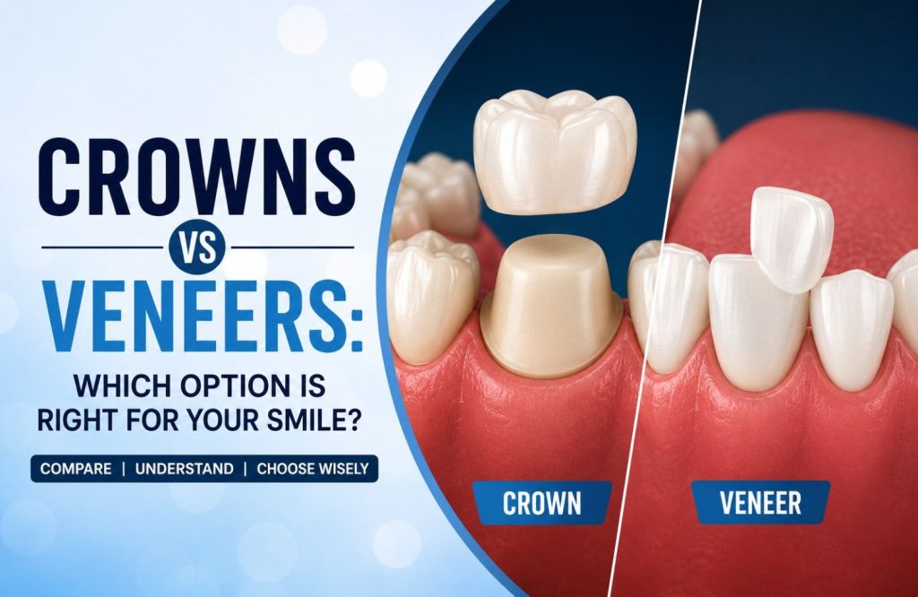
Crowns and veneers come up in the same conversations, recommended by the same dentists, and both change how a tooth looks. That’s about where the similarity ends. What separates them isn’t aesthetics. Both of them can produce a convincing, natural result on the right tooth. The difference is what’s happening structurally underneath. How much tooth remains? What it’s been through? And what it needs to keep functioning reliably going forward? Some situations allow a genuine choice between the two. Others don’t, and the tooth’s condition is what decides, not patient preference. Knowing which side of that line you’re on is what changes the conversation. Two Different Solutions to Two Different Problems A crown encases the entire visible portion of the tooth above the gum line. The outer structure is prepared down on all sides and the crown fits over what remains, replacing the tooth’s exterior completely. The root stays. Where enough inner structure survives, that stays too. What changes is everything the eye sees and everything that takes the pressure of biting. A veneer is a thin shell, typically 0.5 to 1mm, bonded to the front-facing surface only. The back of the tooth, the biting edge, the sides remain largely as they are. A veneer changes what’s visible when the tooth faces forward. The structural situation underneath doesn’t change with it. That difference in coverage is what determines when each is used. Crowns are restorative first. Veneers are cosmetic first, and they require the tooth underneath to be sound enough to make that a viable starting point. What Happens to the Tooth Underneath Before anything is fitted, something has to happen to the tooth. That process is where crowns and veneers diverge most clearly. Crown preparation removes structure from all surfaces. Front, back, sides, the biting surface. The tooth is reduced circumferentially to create space for the crown to sit over it without altering the bite. How much comes off depends on the material and the state of the tooth beforehand, but the preparation covers every side. Veneer preparation removes a thin layer from the front surface only, typically under 1mm. Everything else stays as it is. Ultra-thin veneers sometimes require even less, occasionally none at all, depending on the tooth’s existing dimensions. A tooth that has lost significant structure to decay, fracture, or repeated filling work doesn’t offer enough sound surface for a veneer to bond to reliably. Veneer preparation assumes a largely intact tooth as its starting point. When that starting point is gone, full coverage isn’t a stylistic decision. The less natural tooth structure that remains, the more a crown becomes the clinically correct choice rather than an alternative one. Both treatments follow the same two-appointment sequence: preparation and impressions first, fitting the final restoration second. A temporary restoration protects the tooth between visits. When the Tooth’s Condition Makes the Decision For many patients, the decision has already been made before they sit in the chair. The tooth’s condition is what decided it. A crown is the appropriate treatment when (and the different types of dental crowns available will depend on your specific case): The tooth has extensive decay. Once decay has consumed a substantial portion of the structure, what remains after the cavity is cleared isn’t enough surface for a veneer to bond to reliably. The crown restores what’s missing and protects what’s left. The tooth is cracked. Cracks that compromise structural integrity need full coverage to prevent the fracture from spreading under biting pressure. A veneer bonded to a cracked tooth doesn’t address what’s happening beneath the surface. The tooth has had root canal treatment. Removing the pulp leaves the tooth without its internal moisture source. Root canal treated teeth become brittle over time and fracture under loads that a healthy tooth would handle without issue. Full crown coverage distributes that force and protects against it. A root canal treated tooth left without a crown is one of the more common causes of avoidable tooth loss. A large filling occupies more of the tooth than the natural structure around it. The remaining walls are thin. A crown holds them together. A veneer relies on them to hold it. A cusp has fractured. The damage changes how load is distributed across the whole tooth, not just the visible surface. Front coverage alone doesn’t resolve that. Placing a veneer over a tooth in any of these situations doesn’t fix the problem. When the tooth eventually fails under it, the situation is usually harder to resolve than it would have been with a crown from the start. When the Goal Is Cosmetic and the Tooth Is Sound When the tooth underneath is structurally intact, a crown takes away more than the situation calls for. The preparation required for full coverage on a healthy tooth removes structure that didn’t need to go. In these cases a veneer is the appropriate choice, and it’s chosen because the tooth warrants it, not because it’s the easier option. If you’re considering getting veneers, this is where they genuinely make sense. Veneers are the appropriate treatment when: The tooth is permanently discolored and whitening hasn’t reached it. Tetracycline staining, fluorosis, and trauma-related discoloration sit within the tooth structure rather than on its surface. A veneer covers it fully without altering the structural integrity of the tooth underneath. The tooth is chipped or mildly worn. Where the damage is cosmetic and the structure beneath is sound, a veneer restores what’s visible without the circumferential preparation a crown requires. The tooth is slightly misshapen or disproportionate. Irregularities in length, shape, or surface texture on front teeth that are otherwise healthy are precisely what veneers address. Small gaps or spacing issues exist between front teeth. Where the bite is sound and the concern is cosmetic, veneers can close or reduce spacing in the right cases without orthodontic treatment. Several front teeth need a unified cosmetic result. Veneers placed across the smile line can align the color, shape, and proportion of multiple teeth on a patient whose underlying dentition is healthy enough to support them. On a sound tooth, preserving natural structure is always the better clinical outcome. A veneer does that. A crown on the same tooth wouldn’t. What Patients Usually Ask Before Deciding Does getting a veneer damage the tooth underneath? Veneer preparation removes a thin layer of enamel from the front surface permanently. Enamel doesn’t regenerate, so the tooth will always need a veneer or crown covering that surface from that point forward. That’s the part worth understanding before committing to the treatment. It isn’t structural damage. The dentin, pulp, and root aren’t involved in the preparation. What
Is it tooth sensitivity or somethings else
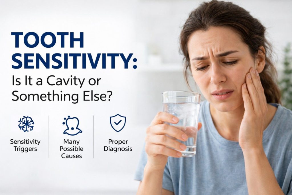
Cold water hits one tooth and the reaction disappears before it fully registers. A few hours later, something sweet triggers the same spot again. Then nothing for the rest of the day. That inconsistency is what makes sensitivity difficult to read properly. Cavities can cause it, but so can exposed roots, enamel wear, grinding, even brushing habits that have been repeated the same way for years. For everything about tooth sensitivity, it helps to understand the full picture first. Sometimes the tooth settles completely after a few seconds. Sometimes the sensation lingers longer than it used to, or keeps returning to the same area. The feeling itself rarely explains much on its own. What’s Actually Happening When a Tooth Feels Sensitive Under the enamel sits a softer layer called dentin, threaded with microscopic channels that lead to the nerve. Enamel keeps them sealed. When it wears down or gums recede, those channels open up to cold, heat, sugar, pressure. The nerve picks up all of it. That response feels identical whether the cause is decay, grinding, gum recession, or acid erosion. The sensation gives you nothing to go on. What’s causing it is a different question entirely, and the answer is what changes the treatment. Not Every Sensitive Tooth Has a Cavity Behind It Sensitivity is the symptom. A cavity is one possible cause. Patients often arrive convinced they’re the same thing, and that assumption tends to create a lot of anxiety before anything has actually been looked at. Several conditions produce sensitivity with no decay involved: Gum recession exposes the root surface, which has no enamel. Root surfaces react to temperature and pressure far more readily than the crown of the tooth. Enamel erosion from acidic food and drinks strips the protective layer gradually. Citrus, fizzy drinks, coffee. It happens slowly enough that sensitivity is often the first sign something has changed. Bruxism wears enamel down through grinding or clenching, frequently during sleep. A significant number of patients find out they’re doing it only after the damage shows up. Brushing too hard with a firm-bristled brush damages both enamel and the gum margin. It’s a slow process, which makes it easy to overlook. A cracked tooth produces sensitivity that can be nearly indistinguishable from a cavity: sharp, localized, triggered by temperature or biting pressure on a specific spot. Recent dental treatment, cleaning, whitening, a new filling, can leave teeth reactive for days to a couple of weeks. This is temporary and resolves on its own. The pattern behind the sensitivity matters as much as the sensitivity itself. Recession tends to affect multiple teeth near the gum line. Bruxism usually shows up across the back teeth. A crack almost always points to one tooth, one spot, one specific trigger. These distinctions are what a clinical assessment is actually looking for. When Sensitivity Is Trying to Tell You Something More Serious The distinction between general sensitivity and a cavity isn’t always clean, but certain signals shift the picture toward decay. Duration is one of the clearest. Sensitivity from worn enamel or gum recession spikes and disappears within seconds of removing the trigger. Cavity-related sensitivity tends to linger; the trigger sets it off, but the discomfort stays for a minute or more after the cause is gone. That difference in duration reflects how deeply the irritation has reached. Location narrows things further. Sensitivity spread across several teeth points toward something systemic, diet, grinding, brushing habit. The same tooth, the same spot, every time, is a different conversation. Sweet foods are a more specific signal than most people expect. Temperature sensitivity can come from several non-cavity sources. Sensitivity reliably triggered by sugar, particularly concentrated in one tooth, correlates more closely with early decay. The bacteria behind cavities produce acid as a byproduct, and that reaction to sugar is fairly distinct. A few other things worth noting: Pain when biting down, separate from temperature or sweetness, suggests structural involvement. Sensitivity that has been gradually worsening over weeks, not staying stable. A visible dark spot, a rough surface, or a texture that feels different than it used to. Discomfort that appears without any trigger, or that wakes you up. Any one of these on its own might mean little. Several of them together, or one that keeps returning, is worth having looked at sooner rather than later. The Pattern of Your Sensitivity Matters More Than the Sensation Itself Two patients can describe identical sensitivity and be dealing with completely different problems. The sensation itself carries less information than when it happens, how long it stays, which teeth are involved, and whether it’s changing over time. A few things worth paying attention to before an appointment: How long does it last? Sensitivity that clears within 30 seconds of removing the trigger points to surface involvement. Lingering past a minute means the nerve is closer to what’s happening. One tooth or several? Sensitivity across multiple teeth suggests something broad, acid erosion, grinding, brushing damage. The same tooth every time is a more specific finding. Is it getting worse? Sensitivity that appeared once and hasn’t returned is different from something that started mild a few weeks ago and has been quietly building. How it changes over time matters more than how it feels on any given day. What sets it off? Temperature sensitivity is common across many causes. Sweet-triggered sensitivity in one specific tooth is a narrower signal. Pain from biting pressure points toward structural damage. Dentists ask these questions because a visual exam doesn’t always show the full picture. Cavities forming between teeth or below the gum line stay hidden until they’re well established. The pattern often gets there first. What a Dentist Is Actually Looking For Before anything is examined, there are questions. When the sensitivity started, what brings it on, whether it’s one tooth or spread across several, whether it’s been shifting. That conversation does more diagnostic work than most patients expect, often before a single instrument is picked up. The examination fills in what the history can’t show. Gums and tooth surfaces checked visually, probing for recession or wear, the bite assessed for uneven pressure. If the clinical picture suggests decay between teeth or below the gum line, where nothing appears on the surface, X-rays follow. An early cavity usually means a dental filling and not much else. Caught before it reaches the nerve, it stays a contained procedure. Left another six months, the same cavity rarely