Crowns and bridges don’t fail the way a toothache announces decay. Some failure modes are immediate and obvious. Others develop over months without producing a symptom the patient connects to the restoration until the problem is considerably more advanced than it needed to be.
The difference between catching a failing crown early and catching it late isn’t just clinical. It’s the difference between a straightforward replacement and a situation where what’s left of the underlying tooth has changed the options available. Secondary decay, cement failure, structural damage, and biological changes beneath the restoration all follow a trajectory. Where a patient is on that trajectory when they seek assessment determines how the situation resolves.
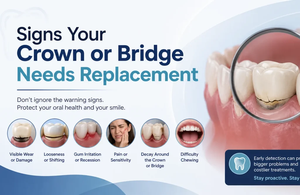
What follows is a specific account of the signs that indicate a crown or bridge needs attention, what each one means, and how urgently it warrants assessment rather than continued monitoring.
When a Crown That Didn’t Hurt Before Starts To
A crown that was comfortable for years and has started producing sensitivity or pain is telling you something specific. What it’s telling you depends on whether the tooth beneath it has a nerve.
Vital teeth under crowns retain their nerve supply. Some temperature sensitivity is possible if the crown margin has degraded and exposed dentinal surfaces near the gum line. Brief sensitivity that clears within seconds of removing the stimulus, unchanged in character and frequency over time, can be assessed at the next routine appointment. Sensitivity that lingers after the stimulus is gone, or that has been gradually increasing, is a different clinical situation. The distinction between the two matters because one is a stable finding and the other is a progressive one.
Sensitivity specifically to biting pressure in a previously comfortable crown narrows the picture. It can reflect a change in the bite relationship, which is adjustable, but it can also reflect secondary decay in the underlying tooth or early seal failure at the crown margin. A bite adjustment and secondary decay produce the same biting sensitivity from the patient’s side. X-ray and clinical examination distinguish between them, which is why the symptom warrants assessment rather than continued monitoring.
A root-canal-treated tooth has no nerve supply. A crown over that tooth shouldn’t produce spontaneous pain. When it does, the source is at or beyond the root tip: periapical pathology from a failing root canal, a root fracture running beneath the crown, or bone involvement in the surrounding tissue. None of those resolve without intervention. The window for the less invasive options, root canal retreatment rather than extraction, narrows as pathology progresses. Pain in a non-vital crowned tooth warrants prompt assessment rather than a watch-and-see approach.
Aching without a clear trigger, particularly if it persists beyond any identifiable stimulus or occurs at rest, points toward a biological cause rather than a mechanical one. That character of discomfort, in any crowned tooth, moves the urgency level up regardless of other factors.
A Crown That Moves Is a Crown That’s Failing
Crown mobility has a specific clinical meaning. A crown that can be felt moving has lost its seal against the prepared tooth beneath it, and the space that creates is the problem rather than the movement itself.
Cement washout is the most common cause. The luting cement bonding the crown to the tooth dissolves gradually, particularly in patients with higher salivary acidity or in older restorations where the original cement layer was thin. The dissolution produces no symptoms until the gap is large enough to produce detectable movement. By that point the seal has already been breached for some time.
The gap between a loose crown and the tooth beneath it is warm, moist, and unreachable by brushing. Bacteria colonize it quickly. Decay in that environment progresses faster than on an open tooth surface because the crown holds the bacterial environment against the tooth while blocking salivary access. A crown recemented weeks after mobility began, without first assessing the tooth beneath, risks sealing active decay in place.
Mobility that has been present for several weeks isn’t the same clinical situation as a crown that came off yesterday. The longer the gap has existed, the more likely it is that the tooth structure available for a new restoration has been affected. Prompt assessment after noticing mobility preserves more options than waiting until the crown falls off on its own.
Over-the-counter dental cement has a specific and limited role: temporary protection when a crown comes off completely and an appointment isn’t immediately available. A crown that is moving but still in place isn’t a situation that temporary cement addresses. It warrants an appointment within days.
The Problem That Develops Without Announcing Itself
A crown protects what it covers. The margin where it meets the tooth at the gum line is where that protection ends, and in many crown failures, that’s where the problem begins.
Bacteria access the margin through micro-gaps that develop as cement ages or as the margin fit degrades over years. Gum recession that exposes a margin previously sitting below the tissue level opens new access points. Once bacteria establish at the margin they work inward along the tooth surface beneath the crown. The crown above remains intact. Nothing at the surface changes until the decay beneath it has progressed far enough to affect the crown’s fit, compromise the underlying structure, or produce a symptom.
The symptom that surfaces most specifically is a persistent bad taste or smell near the crown that brushing doesn’t resolve. Patients who experience this tend to attribute it to food trapping or gum irritation. The distinction is persistence: a food trap produces a transient taste that clears with cleaning. A taste that returns to the same location daily, despite consistent oral hygiene, points toward bacterial activity at the crown margin or within developing decay beneath it. It’s one of the few surface signs of subsurface decay that the patient can identify before clinical examination confirms it.
Visible darkening at the crown margin, a grey or brown line where the crown meets the tooth or gum, can indicate staining at a degraded margin or the shadow of subsurface decay. It isn’t diagnostic on its own. Combined with the persistent taste or any sensitivity, it narrows the picture considerably.
X-rays identify secondary decay beneath a crown before it produces symptoms in most cases, which is the primary clinical reason routine check-ups with radiographic assessment matter for patients with existing restorations. Decay identified at an early stage leaves the tooth structure needed for a straightforward replacement. Decay identified after it has reached the pulp means root canal treatment before the new crown goes on. Decay identified after it has destroyed the majority of what the crown was sitting on changes the question from which restoration to whether restoration is still possible.
Cracks, Chips, and Wear the Eye Can See
Not all visible crown damage carries the same clinical weight. A chip that looks significant may be a cosmetic finding. A crack that looks minor may have broken the crown’s seal in a way that matters considerably more. Location and what the damage affects are the relevant frame, not how it looks.
A chip on the biting surface of a ceramic crown that hasn’t altered the fit, changed the bite, or reached toward the crown margin is primarily cosmetic. In a posterior position, it can be assessed at the next routine appointment. In an anterior position where it’s visible in every interaction, the urgency is driven by the patient’s experience of the result rather than by biological risk.
A crack running through the crown material rather than across a surface layer raises a different question: has it reached the margin? A crack confined to the outer ceramic layer without extending to the margin is a localized structural failure. A crack that reaches or passes through the margin means the seal between the crown and the tooth has been compromised. Bacteria access that gap the same way they access the gap around a loose crown. The crack may look small. The margin breach is the finding that matters.
Significant wear that has flattened the biting surface and changed how upper and lower teeth meet creates a problem beyond the crown. Altered force distribution affects adjacent and opposing teeth over time. In patients with bruxism who aren’t using a night guard, crown wear is frequently where the cumulative effect of parafunctional loading first becomes visible. The crown is showing what’s been happening to the whole dentition.
A crown worn through to its substructure, metal becoming visible through thinning ceramic in a PFM or layered zirconia restoration, has reached a replacement indication regardless of whether it feels structurally sound. The exposed substructure changes how the surface wears against opposing teeth and removes the aesthetic function the crown was placed to provide.
Partial separation, where one side of the crown has lifted from the tooth while the other remains seated, is a same-week assessment situation. The margin is open. The tooth beneath it is no longer protected.
What Bridge Problems Look Like That Crown Problems Don’t
A bridge differs from a single crown in one clinically significant way: the components are interdependent. Two abutment teeth anchor a suspended pontic across the gap left by the missing tooth. A problem at either abutment affects the whole structure. A problem beneath the pontic affects the tissue and bone that any future restoration in that area will depend on.
Food trapping beneath the bridge pontic is the sign most bridge patients notice first and the one most commonly dismissed as a cleaning inconvenience. When a bridge is placed, the pontic sits close to the gum ridge beneath it. As gingival tissue recedes over years, a gap opens between the pontic and the ridge. Food collects there during eating and is difficult to remove because the bridge structure sits above the gap. The trapping reflects a tissue change that creates conditions for bacterial accumulation in an area the patient cannot fully clean. Unmanaged, it progresses to periodontal deterioration in the underlying ridge, which matters for what options exist if the bridge eventually needs replacing.
Sensitivity or pain in one of the abutment teeth follows the same assessment logic as pain in a single crowned tooth, with the additional weight that the abutment tooth’s condition determines whether the bridge remains viable. Abutment teeth carry the functional load of the missing tooth on top of their own. That compounded stress over years produces the same failure modes as any single crown: secondary decay, cement failure, or periapical pathology in a root-canal-treated abutment. A problem in one abutment isn’t just a problem with that tooth.
A rocking sensation when biting on a bridge suggests at least one abutment has lost its cement seal. The lever mechanics of a bridge mean that an unsealed abutment at one end causes the opposite end to lift under load. The mobility the patient feels may be at one location. The cement failure may be at the other. Clinical examination establishes which abutment has failed and whether recementation is appropriate or whether the bridge as a whole has reached the end of its functional life.
Visible darkening or decay at either abutment margin warrants the same prompt assessment as in a single crown, with the understanding that abutment tooth integrity is the foundation the entire bridge rests on. Decay that progresses to compromise an abutment structurally doesn’t just affect that tooth. It determines what replaces the bridge and whether the gap can be bridged again or requires a different solution entirely.
Swelling or tenderness beneath the pontic, in the gum ridge the bridge spans, indicates bacterial accumulation in a space the patient can reach only partially. Left unmanaged, it progresses to bone loss in that ridge. Bone loss in a potential implant site isn’t irreversible, but it adds complexity and cost to what the restoration options look like if the bridge eventually fails.
The Clinical Consequences of Leaving a Failing Restoration in Place
Each failure mode covered in this blog has a clinical trajectory. Where a patient sits on that trajectory when they seek assessment determines what the options look like. The options at early detection are consistently simpler than the options after the problem has progressed, and the cost difference between the two points follows the same direction.
A loose crown left in place for weeks rather than days accumulates secondary decay in the gap between the crown and the tooth at a rate faster than decay on an open surface. Salivary access is blocked. The bacterial environment is warm and moist and undisturbed. A crown recemented over developing decay doesn’t reverse the process. It continues beneath the reseated crown until it produces a symptom, at which point the treatment required has moved from recementation to root canal treatment and a new crown, or beyond that to extraction if enough structure has been lost.
A crack reaching the crown margin and left unaddressed gives bacteria consistent access to the tooth beneath. In a vital tooth, that leads to decay and eventual pulpal involvement. In a root-canal-treated tooth, a crack that extends into the root converts what would have been a crown replacement into an extraction decision. Root fractures beneath crowns are frequently asymptomatic as they propagate. The patient notices nothing. The fracture progresses. By the time a symptom appears, the root can no longer be retained, and the restoration conversation shifts from a new crown to an implant or bridge.
Periapical pathology beneath a root-canal-treated crown spreads to surrounding bone if left untreated. Bone loss from an untreated periapical abscess complicates both retreatment options and any implant placement that might follow extraction. An implant into a site with insufficient bone volume requires grafting before it can be placed. The grafting requirement traces back to a failing root canal that wasn’t assessed when the crown started producing symptoms.
Secondary decay identified before it reaches the pulp means crown removal and a new restoration on remaining structure. The same decay after pulpal involvement means root canal treatment first. After it has destroyed the majority of the coronal structure, the question is whether a post-and-core build-up is possible or whether extraction is the remaining option. The clinical distance between those three points is determined by how long assessment was delayed after the first sign appeared.
What a Crown or Bridge Assessment Actually Involves
A crown or bridge assessment follows a specific and predictable clinical sequence. Visual examination of the restoration and surrounding tissue. Gentle probing at the crown margin to assess seal integrity and detect any gap between the crown and the tooth. Mobility testing. Bite assessment. Targeted periapical X-rays of the tooth or teeth involved, which show what’s happening at the root tip, beneath the crown margin, and in the surrounding bone that the surface examination can’t access.
The combination of clinical and radiographic findings produces a specific picture rather than a general impression. A loose crown with no secondary decay beneath it is a different clinical situation from a loose crown sitting over active caries. A surface chip that hasn’t reached the margin is a different finding from a crack that has compromised the seal. The treatment recommendation follows from what the examination establishes, not from the symptom that prompted the appointment.
When replacement is indicated, the material decision that follows depends on the tooth’s position, the bite load it carries, and the patient’s habits. The clinical reasoning behind crown material selection, and what determines the appropriate choice for a specific tooth and clinical situation, is covered in detail in Different Types of Dental Crowns Explained.
A dental crown and bridge assessment in Abu Dhabi moves the question from what might be happening to what is actually happening. One appointment produces that answer.